Craniopharyngioma Treatment: Craniopharyngiomas are rare, benign brain tumors that typically arise near the pituitary gland, a small organ at the base of the brain responsible for hormone production.
Despite their non-cancerous nature, craniopharyngiomas can significantly impact an individual’s quality of life due to their location and the critical functions of the surrounding brain structures.
This comprehensive guide provides in-depth information on the diagnosis and treatment of craniopharyngiomas, aiming to enlighten patients, caregivers, and healthcare professionals alike.
What is Craniopharyngioma?
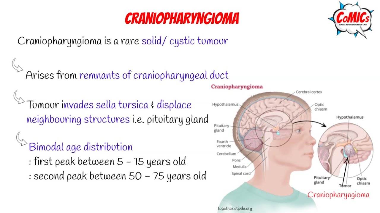
Craniopharyngioma is a rare type of noncancerous (benign) brain tumor that typically occurs near the pituitary gland, a small endocrine gland at the base of the brain. This gland plays a crucial role in regulating vital body functions and the hormonal system. Despite its benign nature, a craniopharyngioma can affect nearby brain structures, leading to significant health problems due to its location.
Characteristics of Craniopharyngioma
Craniopharyngiomas are known for several key characteristics:
- Location: They primarily develop near the pituitary gland, affecting the hypothalamus, optic nerves, and other critical areas of the brain.
- Type: These tumors are benign, meaning they do not spread to other parts of the body. However, they can grow large and impact nearby brain tissue.
- Symptoms: Symptoms can vary widely but often include headaches, vision problems, hormonal imbalances, growth delays in children, and changes in behavior or personality.
- Treatment Complexity: Due to their sensitive location, treating craniopharyngiomas can be complex, often requiring a multidisciplinary approach that may include surgery, radiation therapy, and hormonal replacement therapy.
Epidemiology: Incidence Rates and Demographic Information
Craniopharyngiomas are rare, with an estimated incidence rate of 1.3 cases per million people per year. They can occur at any age but are most commonly diagnosed in children between the ages of 5 and 14 and in adults between 50 and 74 years old. There is no significant gender predilection, and these tumors are observed worldwide, affecting individuals of all ethnicities.
Causes and Risk Factors
The exact cause of craniopharyngioma remains unknown, which is a common scenario with many types of brain tumors. However, research suggests that genetic factors may play a role in their development. Unlike many other tumors, craniopharyngiomas are not associated with environmental or lifestyle risk factors. Studies have investigated potential genetic mutations that could contribute to tumor formation, but clear, direct causes have yet to be established. This uncertainty highlights the need for ongoing research to better understand the origins of craniopharyngioma and to develop targeted treatments.
However, craniopharyngiomas are rare, benign brain tumors with a complex presentation due to their critical location near the pituitary gland. They can cause a variety of symptoms related to hormonal imbalances and pressure on nearby brain structures. Despite being benign, their treatment requires careful consideration due to the delicate area of the brain they affect. Current understanding of their causes is limited, emphasizing the role of genetic factors, though much remains to be discovered about why these tumors develop.
Symptoms of Craniopharyngioma: An In-depth Analysis
Due to its critical location, the presence of a craniopharyngioma can lead to a variety of symptoms that not only affect physical health but can also impact an individual’s quality of life. Understanding these symptoms and their underlying causes is crucial for early detection and effective management of the condition.
Detailed List of Symptoms
The symptoms of craniopharyngioma can be diverse, reflecting the tumor’s impact on the brain’s functioning and its pressure on nearby structures. Key symptoms include:
- Headaches – Often the first noticeable sign, caused by increased pressure within the skull.
- Vision Problems – Including blurred vision, double vision, or loss of peripheral vision due to the tumor pressing on the optic nerves.
- Hormonal Imbalances – Manifesting as growth delays in children, irregular menstrual cycles in women, increased thirst and urination, and unexpected weight gain or loss, due to the tumor’s effect on the pituitary gland.
- Fatigue – A common symptom resulting from hormonal imbalances affecting the body’s energy levels.
- Mood Swings and Behavioral Changes – Resulting from the tumor’s pressure on brain areas that regulate mood and behavior.
- Nausea and Vomiting – Typically occurring in the morning and worsening with time, related to increased pressure within the brain.
Why These Symptoms Occur
The symptoms of craniopharyngioma are closely linked to the tumor’s location and its impact on surrounding brain structures and the pituitary gland. The pituitary gland, often termed the “master gland,” controls a multitude of hormones that regulate vital body functions, including growth, metabolism, and reproductive processes. When a craniopharyngioma develops, it can press on the pituitary gland and adjacent parts of the brain, disrupting their normal functioning.
- Impact on Vision: The optic chiasm, a critical structure for vision, is located near the pituitary gland. As the tumor grows, it can exert pressure on this area, leading to visual disturbances.
- Hormonal Dysfunction: Given the pituitary gland’s role in hormone production, a craniopharyngioma can significantly disrupt its function. This disruption can lead to an imbalance in hormone levels, affecting the body’s normal operations and leading to symptoms like growth delays or menstrual irregularities.
- Increased Intracranial Pressure: As the tumor enlarges, it can increase pressure inside the skull, contributing to headaches, nausea, and cognitive changes.
If you or someone you know is experiencing these symptoms, consulting with a healthcare professional for a thorough evaluation is advised. Understanding the intricate relationship between the tumor’s location and its symptoms is essential for grasping the full scope of this condition’s effects on the body.
Diagnosis of Craniopharyngioma
Effective diagnosis involves a series of steps, incorporating advanced imaging techniques and, in certain cases, biopsy. This article outlines the diagnostic process for craniopharyngioma and delves into the crucial role of biopsy in confirming the diagnosis.
Diagnostic Process for Craniopharyngioma
The journey to a definitive diagnosis of craniopharyngioma typically involves the following steps:
Medical History and Physical Examination: The first step in diagnosing craniopharyngioma involves a thorough review of the patient’s medical history and a physical examination. Doctors look for symptoms common to craniopharyngioma, such as headaches, vision problems, hormonal imbalances, and growth issues in children.
Imaging Tests:
- MRI (Magnetic Resonance Imaging): An MRI is pivotal in the diagnosis of craniopharyngioma. It provides detailed images of the brain, highlighting the tumor’s size, location, and impact on surrounding structures.
- CT (Computed Tomography) Scan: Though less detailed than MRI, a CT scan can help identify calcifications within the tumor, a characteristic feature of craniopharyngioma.
Hormonal Testing: Since craniopharyngiomas often affect the pituitary gland, hormonal testing is crucial. These tests assess the levels of various hormones in the blood to identify any imbalances caused by the tumor.
Vision Tests: Given the tumor’s potential to impact the optic nerves, vision tests are conducted to assess any loss of vision or changes in field of vision.
Role of Biopsy in Diagnosis
While imaging tests and hormonal assessments provide substantial information, a biopsy is the definitive method for diagnosing craniopharyngioma. However, it’s not always the first step due to the risks involved with brain surgery. The role of biopsy in diagnosing craniopharyngioma includes:
- Confirming the Diagnosis: A biopsy involves removing a small sample of the tumor tissue. This sample is then examined under a microscope to confirm the presence of craniopharyngioma cells.
- Determining the Type: Craniopharyngiomas can be classified into two types based on their histological characteristics: adamantinomatous and papillary. A biopsy helps in determining the tumor type, which is crucial for planning the treatment.
When is Biopsy Used? Biopsy is typically considered when:
- The imaging tests are inconclusive.
- The tumor’s location allows for a safe biopsy procedure.
- A definitive diagnosis is required to guide treatment decisions, especially if the treatment approach may vary significantly depending on the tumor type.
However, the diagnosis of craniopharyngioma is a multifaceted process that involves a combination of medical history review, physical examination, imaging tests, hormonal testing, and vision tests. Biopsy plays a critical role, especially in cases where the diagnosis remains uncertain after initial tests. Despite the complexities involved, accurate diagnosis is crucial for effective treatment and management of craniopharyngioma.
Treatment Options for Craniopharyngioma
Here, we explore the main treatment avenues including surgical treatment, radiation therapy, medication and hormone replacement therapy, and look into emerging treatments and research that offer hope for the future.
Surgical Treatment
Surgery is often the first line of treatment for craniopharyngioma. The goal is to remove as much of the tumor as possible without damaging the surrounding brain tissue. Advances in surgical techniques, such as the use of microscopes and endoscopes, have significantly increased the safety and effectiveness of these operations. There are two main surgical approaches:
- Transsphenoidal surgery: This less invasive approach is used for tumors that are located entirely or mostly in the pituitary gland or below it. The surgeon accesses the tumor through the nasal cavity, minimizing damage to the brain.
- Craniotomy: For tumors that cannot be reached transsphenoidally, a craniotomy may be necessary. This involves opening the skull to access the tumor directly.
Despite the advancements, surgical treatment carries risks such as damage to surrounding brain structures, infections, and the need for further treatments if the entire tumor cannot be removed.
Radiation Therapy
Radiation therapy is another cornerstone in the treatment of craniopharyngioma, particularly when surgical removal is incomplete or not possible. It involves the use of high-energy beams to target and kill tumor cells. Techniques like stereotactic radiosurgery (SRS) and fractionated stereotactic radiotherapy (FSRT) allow for precise targeting of the tumor, minimizing exposure to healthy brain tissue. Radiation therapy can be effective in controlling tumor growth, but it also comes with potential side effects, including effects on cognitive function, growth in children, and the risk of secondary tumors.
Medication and Hormone Replacement Therapy
Due to the tumor’s proximity to the pituitary gland, hormone deficiencies are a common complication. Medication and hormone replacement therapy play a critical role in managing these deficiencies and maintaining normal body functions. Patients may require lifelong hormone replacement therapy for hormones such as cortisol, thyroid hormone, growth hormone, and sex hormones. Additionally, medications may be used to manage symptoms or complications related to the tumor or its treatment.
Emerging Treatments and Research
The future looks promising with ongoing research and emerging treatments for craniopharyngioma. Targeted therapy, which aims at specific genes, proteins, or the tissue environment that contributes to tumor growth, is under investigation. Immunotherapy, a treatment that uses the body’s immune system to fight cancer, is another area of active research. Clinical trials are also exploring novel approaches to improve outcomes and reduce side effects. These emerging treatments could offer new hope for patients with craniopharyngioma, especially those for whom traditional treatments are not effective.
However, the treatment of craniopharyngioma involves a multidisciplinary approach that includes surgical intervention, radiation therapy, medication, and hormone replacement therapy. With ongoing research and the development of new treatment modalities, the outlook for patients with craniopharyngioma continues to improve. It is essential for patients and their families to work closely with their healthcare team to choose the best treatment plan tailored to their specific needs.
Living with Craniopharyngioma
Living with craniopharyngioma, a rare type of brain tumor that typically affects children and adults, involves navigating a variety of long-term effects and complications. This journey requires not only medical intervention but also a comprehensive support system to enhance the quality of life for those affected. Understanding the importance of regular follow-up care, supportive services, and rehabilitation is crucial for patients and their families.
Managing Long-Term Effects and Complications
After treatment for craniopharyngioma, which may include surgery, radiation therapy, or a combination of treatments, patients often face long-term effects and complications. These can range from vision problems and hormonal imbalances to neurological deficits. It’s essential to manage these effects proactively:
- Hormonal Imbalances: Many patients experience issues with their pituitary gland, which can affect hormone production. Regular monitoring and hormone replacement therapy can help manage these imbalances.
- Vision Problems: Given the tumor’s proximity to the optic nerves, vision issues are common. Regular eye exams and consultations with an ophthalmologist can help detect and manage any changes in vision.
- Neurological Issues: Some patients may experience cognitive or physical impairments. Working with neurologists and rehabilitation specialists can help maximize neurological function and independence.
Importance of Regular Follow-Up Care
Regular follow-up care is paramount for individuals living with craniopharyngioma. These appointments allow healthcare providers to monitor for tumor recurrence, a crucial step since craniopharyngiomas can recur even after successful treatment. Additionally, follow-up visits are critical for managing endocrine disorders that often result from the tumor or its treatment. Endocrinologists play a vital role in adjusting hormone replacement therapies as needed to maintain hormonal balance and overall health.
Supportive Care and Rehabilitation Services
Supportive care and rehabilitation services are indispensable for improving the quality of life for craniopharyngioma patients. These services address the physical, emotional, and social challenges that can arise:
- Physical Rehabilitation: Physical therapists can help patients regain strength and mobility, improving independence and functionality.
- Psychological Support: Mental health professionals can provide counseling to help patients and families cope with the emotional impact of the diagnosis and its aftermath.
- Social Services: Social workers can assist families in navigating the healthcare system, accessing community resources, and securing financial assistance if needed.
By managing long-term effects and complications, ensuring regular follow-up care, and utilizing supportive care and rehabilitation services, patients can navigate the challenges of this condition. A proactive and supportive approach can significantly enhance the quality of life for those affected by craniopharyngioma, fostering resilience and well-being throughout their journey.
Conclusion
The journey through understanding, diagnosing, and treating Craniopharyngioma underscores the paramount importance of early detection and the implementation of an effective treatment strategy. Recognizing the signs and symptoms of Craniopharyngioma as early as possible is crucial. A timely diagnosis can significantly enhance the prognosis and quality of life for those affected by this complex condition. It opens the door to a broader range of treatment options, including surgical intervention, radiation therapy, and hormone replacement therapy, each tailored to meet the unique needs of the individual patient.
Equally important is the collaborative effort between patients, families, and healthcare professionals in crafting a personalized treatment plan. This team-based approach ensures that all aspects of the patient’s well-being are considered, including physical health, emotional support, and the management of potential side effects or complications. Patients and families are encouraged to actively engage with their medical team, ask questions, and express their concerns and preferences. This open line of communication fosters a supportive environment that is conducive to healing and recovery.
In conclusion, the fight against Craniopharyngioma is a testament to the resilience of patients, the dedication of families, and the expertise of healthcare professionals. Together, through timely diagnosis and customized treatment plans, it is possible to manage this condition effectively, improving outcomes and enhancing the lives of those affected. Let this be a call to action for patients and families to work hand in hand with their healthcare team, navigating the challenges of Craniopharyngioma with courage and hope.