Craniosynostosis Symptoms: Craniosynostosis is a medical condition marked by the premature fusion of one or more cranial sutures in infants, leading to abnormal skull shape and potential developmental issues.
This comprehensive guide delves into the symptoms, causes, and critical aspects of craniosynostosis, offering essential insights for parents and caregivers.
What is Craniosynostosis?
Craniosynostosis is a medical condition characterized by the premature fusion of one or more of the cranial sutures in infants. These sutures are the fibrous joints between the bones of the skull, which typically remain flexible to allow the brain to grow during the early years of life. When these sutures close too early, it can restrict the growth of the brain and skull, leading to potential developmental issues and changes in the shape of the head. Early diagnosis and treatment are crucial in managing the condition and preventing long-term complications.
The Role of Sutures in Skull Development
Sutures play a critical role in skull development by allowing the skull to expand as the brain grows. This flexibility is essential for accommodating the rapid brain growth that occurs during the first few years of a child’s life. The sutures gradually harden over time, usually completing the process by the age of 2 to 3 years. However, when these sutures fuse prematurely, it disrupts normal skull and brain growth. Understanding the role of sutures in skull development is key to comprehending the impacts of craniosynostosis and the importance of early intervention.
Statistics on Prevalence and Demographics
Craniosynostosis is a relatively rare condition, affecting approximately 1 in every 2,500 to 3,000 live births. It can occur as an isolated condition or as part of a genetic syndrome. While it can affect any child, some studies suggest a slightly higher incidence in males compared to females. The condition is also noted to vary by ethnicity and geographical location, though comprehensive global data is limited. Early detection and treatment strategies are critical in managing craniosynostosis effectively, highlighting the importance of awareness and education among parents and healthcare providers.
By understanding what craniosynostosis is, the vital role of sutures in skull development, and the statistics on its prevalence and demographics, parents and caregivers can be better equipped to seek timely medical advice. Early intervention can significantly improve outcomes for children with craniosynostosis, emphasizing the need for awareness and accessible healthcare solutions.
Symptoms of Craniosynostosis
Recognizing the early signs and symptoms of this condition is crucial for timely intervention and treatment. This article outlines the key symptoms of craniosynostosis, how it affects a child’s appearance and health, the variations in symptoms among its types, and when to seek medical advice.
Early Signs and Symptoms to Watch For
The symptoms of craniosynostosis may vary depending on which cranial sutures are affected. However, some common early signs include:
- Misshapen skull: One of the most noticeable signs is an abnormally shaped head, which can be elongated, flattened, or asymmetric.
- A hard, raised ridge along the affected sutures: As the sutures fuse prematurely, a noticeable ridge can form along these areas.
- Slow or no growth of the head as the baby grows: Unlike typical development, a child with craniosynostosis may show limited or no increase in head circumference.
- Increased intracranial pressure: In some cases, symptoms such as irritability, excessive sleepiness, or vomiting may indicate increased pressure within the skull.
How Craniosynostosis Affects a Child’s Appearance and Health
Craniosynostosis can significantly impact both the appearance and the health of a child. Appearance-wise, the condition can lead to a noticeably misshapen head, which may affect the child’s facial symmetry and overall head shape. Health-wise, the condition can lead to developmental delays, including cognitive impairments if not treated promptly. Increased intracranial pressure can cause headaches, vision problems, and in severe cases, brain damage.
Differences in Symptoms Among the Types of Craniosynostosis
There are several types of craniosynostosis, named according to the affected sutures:
- Sagittal synostosis results in a long, narrow skull, as it affects the suture along the top of the head.
- Coronal synostosis affects one or both of the coronal sutures that run from ear to ear, leading to a flattened forehead and brow.
- Metopic synostosis affects the suture close to the forehead, resulting in a triangularly shaped forehead and a ridge down the middle of the forehead.
- Lambdoid synostosis affects the suture along the back of the head, leading to a flattened back of the head.
Each type of craniosynostosis presents a unique set of symptoms and challenges, making it important to identify the specific type for appropriate treatment.
When to Seek Medical Advice
If you notice any of the early signs of craniosynostosis in your child, such as a misshapen skull or a hard ridge along their head, it’s crucial to seek medical advice promptly. Early diagnosis and treatment can prevent complications related to the condition, such as developmental delays and increased intracranial pressure. Consult with a pediatrician or a specialist in pediatric neurosurgery or craniofacial surgery to discuss your observations and concerns. Early intervention is key to ensuring the best possible outcomes for children with craniosynostosis.
However, understanding the symptoms and implications of craniosynostosis is essential for parents and caregivers. By staying informed and vigilant, you can ensure timely medical intervention, helping your child achieve optimal health and development.
Causes and Risk Factors for Craniosynostosis
Understanding the causes and risk factors is crucial for early diagnosis and intervention. Here, we delve into the genetic and environmental factors, as well as the role of family history and hereditary patterns, that contribute to the development of craniosynostosis.
Genetic Factors Contributing to Craniosynostosis
Genetic mutations play a significant role in craniosynostosis. These mutations can either be inherited or occur spontaneously. There are specific genes known to be associated with craniosynostosis, such as FGFR2, FGFR3, and TWIST1. These genes are crucial for bone development and growth, and mutations in these genes can lead to the premature fusion of skull sutures. Genetic testing can help identify these mutations, which is particularly beneficial for families with a history of craniosynostosis, as it provides a clearer understanding of the risk for future children.
Environmental Factors That May Increase Risk
Environmental factors can also increase the risk of developing craniosynostosis. These factors include maternal smoking, use of fertility treatments, and certain medications during pregnancy. Additionally, nutritional factors, such as maternal obesity and diabetes, have been linked to a higher risk of craniosynostosis in infants. It’s important for expectant mothers to have regular prenatal care, which can help manage these risk factors effectively.
The Role of Family History and Hereditary Patterns
Family history and hereditary patterns significantly influence the risk of craniosynostosis. If a family member has had craniosynostosis, particularly if the condition is associated with a known genetic mutation, the risk of another family member being affected increases. This condition can follow autosomal dominant or recessive inheritance patterns. In families with an autosomal dominant pattern, there is a 50% chance of passing the condition to offspring. Understanding these hereditary patterns can aid in early detection and treatment planning.
However, craniosynostosis results from a complex interplay of genetic and environmental factors, with family history and hereditary patterns playing a pivotal role. Awareness of these causes and risk factors can lead to early diagnosis and more effective treatment, significantly improving outcomes for affected infants. Regular prenatal visits and genetic counseling can provide valuable insights for expecting parents, especially those with a family history of craniosynostosis.
Types of Craniosynostosis: An Overview
This article delves into the four main types of craniosynostosis: sagittal, coronal, metopic, and lambdoid. Understanding the distinctions among these types is crucial for early diagnosis and effective treatment planning.
Sagittal Craniosynostosis
Sagittal craniosynostosis, the most common type, occurs when the sagittal suture—the line that runs from the front to the back of the skull—closes early. This premature fusion prevents the skull from widening naturally, leading to a long, narrow skull shape known as scaphocephaly. Symptoms often include a prominent ridge along the sagittal suture and an increased head circumference due to compensatory growth in the areas of the skull that remain unfused.
Coronal Craniosynostosis
This type involves the premature closure of one or both coronal sutures, which stretch from ear to ear over the top of the skull. When one coronal suture fuses early (unicoronal craniosynostosis), it results in an asymmetrical skull shape, with one side of the forehead appearing flatter than the other (anterior plagiocephaly). Bilateral coronal craniosynostosis, where both sutures close prematurely, leads to a short and wide skull, known as brachycephaly. Symptoms can include asymmetry in the facial features and potentially impaired development of the eye socket on the affected side.
Metopic Craniosynostosis
The metopic suture runs from the top of the forehead down the middle towards the nose. Metopic craniosynostosis leads to trigonocephaly, characterized by a triangular forehead and a pointed scalp. This type of craniosynostosis can cause the eyes to appear closer together and may result in a ridge forming on the forehead. Symptoms also include developmental delays and cognitive challenges in some cases.
Lambdoid Craniosynostosis
Lambdoid craniosynostosis is the least common type and involves the early fusion of the lambdoid suture, located at the back of the skull. This condition can cause the skull to grow asymmetrically, resulting in a condition called posterior plagiocephaly, where one side of the back of the head appears flatter than the other. Symptoms may include tilting of the head and a noticeable difference in ear alignment.
Each type of craniosynostosis affects the skull shape and symptoms differently, making it essential for healthcare providers to accurately diagnose the specific type to tailor treatment effectively. Early intervention is critical to prevent potential complications, including increased intracranial pressure, developmental delays, and cognitive impairments. With advances in surgical techniques and early detection, children with craniosynostosis can lead healthy, fulfilling lives.
Complications Associated with Craniosynostosis
Understanding the potential health issues, impact on brain development and function, along with emotional and psychological effects, is crucial for parents and caregivers.
Potential Health Issues Resulting from Untreated Craniosynostosis
Untreated craniosynostosis can result in a range of health issues. These may include increased intracranial pressure due to the restricted growth of the skull, leading to potential damage to brain tissue and consequent developmental delays. Vision problems are also common, as the abnormal shape of the skull can affect the orbits and, subsequently, eye function. Respiratory issues and hearing loss are other potential complications, stemming from the abnormal growth and development of facial structures and ear canals.
Impact on Brain Development and Function
The brain’s development and function can be significantly impacted by untreated craniosynostosis. The increased intracranial pressure associated with this condition can hinder the brain’s ability to grow and develop normally, potentially leading to cognitive impairments and delays in speech and language skills. In severe cases, this pressure can also cause neurological damage, affecting a child’s motor skills and coordination.
Emotional and Psychological Effects
Beyond the physical complications, craniosynostosis can have profound emotional and psychological effects on affected individuals and their families. Children with visible skull deformities may experience social challenges, including bullying and social isolation, leading to low self-esteem and confidence issues. For parents and caregivers, the diagnosis and management of craniosynostosis can be a source of significant stress and anxiety, affecting their emotional well-being and family dynamics.
Early diagnosis and intervention are key to managing craniosynostosis and minimizing its complications. Surgical treatments can correct the skull’s shape, alleviate increased intracranial pressure, and allow for normal brain development. Supportive therapies, such as speech and physical therapy, can also help address developmental delays. Emotional and psychological support for the child and family is equally important, ensuring a comprehensive approach to treatment and care.
However, while craniosynostosis can lead to various complications, understanding these potential issues allows for better preparedness and management. With timely and appropriate treatment, many children with craniosynostosis can lead healthy, fulfilling lives.
Diagnosis of Craniosynostosis
Understanding the various methods and tools used in diagnosing this condition is crucial for healthcare professionals and parents alike. This article delves into the primary diagnostic approaches, underlines the significance of early detection, and highlights the pivotal role of imaging tests in confirming craniosynostosis.
Diagnostic Methods and Tools
Diagnosis of craniosynostosis typically involves a combination of physical examinations and imaging tests. Here are the key methods and tools employed:
- Physical Examination: Initially, healthcare providers conduct a thorough physical examination of the infant’s head. They look for abnormal head shape and feel the sutures and fontanelles (soft spots) for signs of premature closure.
- Cranial Ultrasound: This non-invasive tool is often used for newborns and young infants, providing a quick look at the cranial sutures to identify any premature fusion.
- Computed Tomography (CT) Scan: A CT scan offers detailed images of the skull, allowing doctors to precisely assess the sutures’ status. It’s considered the gold standard in diagnosing craniosynostosis, despite concerns about radiation exposure.
- Magnetic Resonance Imaging (MRI): MRI can be used to evaluate brain structures and look for associated abnormalities without radiation exposure. However, it’s less commonly used specifically for diagnosing craniosynostosis.
The Importance of Early Detection
Early detection of craniosynostosis is crucial for several reasons. It allows for timely intervention, which can prevent or minimize potential complications such as increased intracranial pressure, developmental delays, and abnormal head shape. Early treatment, often involving surgery, can improve outcomes significantly. Therefore, parents and healthcare providers should be vigilant for signs of this condition, especially in the first few months of an infant’s life.
Role of Imaging Tests in Diagnosis
Imaging tests play a vital role in the diagnosis of craniosynostosis. They not only confirm the presence of premature suture fusion but also help in planning the surgical intervention. A CT scan, in particular, provides detailed insights into the extent of suture fusion and guides the surgical team in correcting the skull’s shape effectively. Moreover, imaging tests can help differentiate craniosynostosis from other conditions that might cause abnormal head shapes, ensuring that the child receives the most appropriate care.
However, the diagnosis of craniosynostosis involves a multifaceted approach, combining physical examination with advanced imaging techniques. The importance of early detection cannot be overstated, as it directly impacts the child’s developmental outcomes and quality of life. Imaging tests, especially CT scans, are indispensable in confirming the diagnosis and facilitating successful treatment planning. For parents and healthcare providers, being informed about these diagnostic tools and the significance of early intervention is key to managing craniosynostosis effectively.
Treatment Options for Craniosynostosis
Understanding the available treatment options, factors influencing the choice of treatment, and expected outcomes can empower parents and caregivers in making informed decisions regarding their child’s care.
Treatment Approaches
1. Surgical Correction: The primary and most common treatment for craniosynostosis is surgical intervention. Surgical techniques can vary, including:
- Endoscopic Surgery: Best suited for infants under 6 months, this minimally invasive procedure involves making small incisions to correct the suture fusion. It typically requires a shorter hospital stay and recovery period.
- Open Cranial Vault Remodeling: For older infants, this traditional surgery involves reshaping the skull and is performed to correct more complex or multiple suture fusions. Although it involves a longer recovery time, it has a high success rate in improving skull shape and brain development.
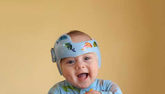
2. Helmet Therapy: Often used in conjunction with surgery, helmet therapy can help mold the skull into a more typical shape as the brain grows. It is most effective when started early and usually involves wearing a custom-fitted helmet for 23 hours a day over several months.
3. Non-Surgical Management: In very mild cases, or when surgery poses too high a risk, careful monitoring may be recommended. This approach is less common and usually involves regular check-ups to monitor the child’s brain development and head growth.
Factors Influencing the Choice of Treatment
Several factors determine the most appropriate treatment approach for craniosynostosis, including:
- Age of the Child: Younger infants may be candidates for less invasive procedures, such as endoscopic surgery.
- Type and Severity of the Craniosynostosis: The specific suture(s) affected and the severity of the fusion influence the surgical approach.
- Overall Health of the Child: Any underlying health conditions can affect the choice of treatment and anesthesia considerations.
- Preference of the Family and Surgical Team: Decisions are made based on a combination of medical advice and the family’s comfort level with the proposed treatment plan.
Expected Outcomes and Prognosis After Treatment
The prognosis for children with craniosynostosis is generally very good, especially when the condition is diagnosed and treated early. Expected outcomes include:
- Improvement in Head Shape: Surgical and helmet therapies can significantly improve the symmetry and shape of the child’s head.
- Normal Brain Development: Early treatment can prevent or minimize potential impacts on brain development, allowing for normal cognitive and physical development.
- Quality of Life: Addressing craniosynostosis early can improve overall quality of life, reducing the risk of psychological and physical complications associated with abnormal head shapes.
However, craniosynostosis treatment is highly effective, particularly when initiated early. Collaboration between healthcare providers and families is crucial to tailor the treatment plan to the child’s needs, ensuring the best possible outcome and quality of life. Regular follow-ups and supportive care play essential roles in monitoring the child’s development and adjusting treatment plans as necessary.
Preventive Measures and Support for Craniosynostosis
While the focus is often on treatment post-diagnosis, understanding preventive measures and the support available to families is crucial. This section delves into the current research on prevention strategies and outlines resources and support systems for families navigating craniosynostosis.
Current Research on Prevention Strategies
Recent advancements in medical research have begun to shed light on potential preventive measures for craniosynostosis. Although genetic factors play a significant role in many cases, making outright prevention challenging, there are areas where ongoing research is promising:
- Maternal Health: Studies suggest that maintaining optimal health during pregnancy may reduce the risk of craniosynostosis. This includes adequate intake of folic acid, proper management of maternal diabetes, and avoiding exposure to harmful substances.
- Genetic Counseling: For families with a history of craniosynostosis, genetic counseling is recommended. It can provide insights into the risk of recurrence in future offspring and discuss possible preventive steps.
- Early Detection: While not preventive per se, early detection of cranial suture abnormalities can lead to timely intervention, potentially mitigating complications. Prenatal imaging and early postnatal assessments are crucial.
Resources and Support for Families Affected by Craniosynostosis
Navigating the journey of craniosynostosis can be challenging for families. However, a robust support system can significantly ease this burden. Here are several resources and forms of support available:
- Specialized Healthcare Teams: Pediatric neurosurgeons, craniofacial surgeons, and pediatricians play vital roles in the treatment and ongoing care of children with craniosynostosis. These professionals not only provide medical care but also guide families through the treatment process.
- Support Groups: Joining a support group for families affected by craniosynostosis can offer emotional support and practical advice. Sharing experiences with others in similar situations can be incredibly comforting.
- Educational Resources: Numerous organizations and websites offer comprehensive information about craniosynostosis. These resources can help families understand the condition, treatment options, and what to expect.
- Financial Assistance Programs: The cost of treatment for craniosynostosis can be high. Some organizations offer financial assistance or guidance on navigating insurance benefits to cover treatment costs.
However, while the prevention of craniosynostosis may not always be possible, understanding potential preventive measures and early detection strategies is essential. Equally important is the availability of resources and support systems for families. By leveraging these supports, families can navigate the complexities of craniosynostosis with greater confidence and less stress, ensuring the best possible outcomes for their children.
FAQs: Common Questions About Craniosynostosis
What is Craniosynostosis?
Craniosynostosis is a condition where one or more of the fibrous joints between the bones of an infant’s skull (cranial sutures) close prematurely, before the brain is fully formed. This can restrict the growth of the skull and affect the shape of the head and face.
What Causes Craniosynostosis?
The exact cause of craniosynostosis is not always known. It can be the result of genetic factors, environmental influences, or a combination of both. Some cases are associated with certain genetic syndromes, but many occur in infants with no family history of the condition.
What Are the Symptoms of Craniosynostosis?
The most noticeable symptom of craniosynostosis is an abnormal shape of the infant’s head. Other symptoms can include a palpable ridge along the sutures, slow or no growth of the head as the infant grows, and in severe cases, increased pressure within the skull.
How Is Craniosynostosis Diagnosed?
Craniosynostosis is typically diagnosed through a physical examination and imaging tests. A doctor may feel the infant’s head for any abnormal ridges and look for facial asymmetry. Imaging tests like X-rays, CT scans, or MRI scans can confirm the diagnosis by showing the fused sutures.
What Are the Treatment Options for Craniosynostosis?
Treatment for craniosynostosis usually involves surgery to correct the shape of the skull and allow for normal brain growth. The type of surgery depends on the sutures involved and the severity of the condition. In some cases, minimally invasive techniques can be used.
Can Craniosynostosis Lead to Complications?
If left untreated, craniosynostosis can lead to complications such as increased intracranial pressure, developmental delays, and visual impairments. Early diagnosis and treatment are crucial to prevent these potential complications.
Is Craniosynostosis Common?
Craniosynostosis is relatively rare, affecting approximately 1 in every 2,500 live births. It can occur as an isolated condition or as part of a syndrome that affects other parts of the body.
Can Craniosynostosis Be Prevented?
Currently, there is no known way to prevent craniosynostosis. However, understanding the risk factors and seeking early medical advice if you notice any signs can help in timely diagnosis and treatment.
Where Can I Find Support?
If your child has been diagnosed with craniosynostosis, it’s important to seek support. Many hospitals and organizations offer resources for families, including counseling, support groups, and educational materials.
Conclusion:
For parents and caregivers, being vigilant and informed about the signs of craniosynostosis is essential. These can range from an unusual head shape at birth to more subtle signs such as developmental delays. It’s crucial to remember that every child’s growth pattern is unique, and variations in head shape are common. However, any concerns regarding your child’s head shape or developmental milestones should prompt a consultation with a healthcare provider.
Seeking medical advice at the earliest sign of concern is paramount. Healthcare professionals can provide comprehensive evaluations to determine if craniosynostosis or another condition is present. Early intervention strategies, which may include surgery, can significantly improve outcomes for children affected by craniosynostosis. These treatments are designed to correct the shape of the skull and ensure proper brain growth and development.
In conclusion, the early detection of craniosynostosis symptoms plays a vital role in the health and development of affected children. Parents and caregivers are encouraged to trust their instincts and seek medical advice for any concerns related to their child’s head shape or developmental progress. Through awareness, early diagnosis, and intervention, we can ensure that children with craniosynostosis receive the care and support they need to thrive. Let’s prioritize the health and well-being of our youngest and most vulnerable by staying informed and proactive in their care.