Craniosynostosis Treatment: Craniosynostosis is a medical condition that affects the skull, leading to premature fusion of one or more cranial sutures in infants.
This early suture closure can result in abnormal skull shape, potential brain growth restriction, and cognitive development issues.
Recognizing the signs and pursuing timely diagnosis and treatment are critical for optimal outcomes.
Understanding Craniosynostosis
Craniosynostosis is a condition that affects the skull’s development in infants and young children, leading to potentially significant health issues and aesthetic concerns. Understanding this condition requires a fundamental grasp of the role sutures play in skull growth and the consequences of their premature fusion. By the end of this section, you will have a clearer understanding of craniosynostosis, including its different types and implications on health.
The Role of Sutures in Skull Growth
The human skull isn’t a single, solid bone but rather a complex structure made up of several bones. These bones are connected by fibrous joints known as sutures. Sutures are crucial in skull growth because they allow the skull to expand as the brain grows, especially during the first few years of life. This expansion is essential for accommodating the growing brain, ensuring that it has enough space to develop properly.
Sutures act like expansion joints, absorbing the growth of the brain and skull. They gradually ossify (turn into bone) over time, a process that is usually complete by the late teens or early twenties. This natural progression allows the skull to increase in size while maintaining its protective function and the brain’s developmental needs.
What Happens When Sutures Fuse Prematurely
Craniosynostosis occurs when one or more of these sutures fuse prematurely, limiting the skull’s growth in specific areas while allowing it to continue expanding in others. This uneven growth can lead to an abnormal head shape and can potentially cause pressure on the growing brain, leading to developmental delays or neurological problems in some cases.
The early fusion of sutures forces the skull to grow in the direction of the remaining open sutures, leading to several possible abnormal shapes. This condition not only affects the shape of the head but can also impact the child’s facial features and, in severe cases, brain development.
Different Types of Craniosynostosis
Craniosynostosis can be categorized into several types, depending on which sutures are affected. The main types include:
- Sagittal Synostosis: The most common form, affecting the suture running along the top of the head from front to back. It leads to a long, narrow skull shape.
- Coronal Synostosis: Involves the premature fusion of one or both of the coronal sutures, which run from ear to ear. It may result in a flattened forehead and brow on the affected side and can cause the eye socket to be elevated, leading to asymmetry.
- Metopic Synostosis: Affects the suture that runs from the top of the forehead down the middle of the forehead to the nose. It can lead to a triangular forehead and a ridge down the center of the forehead.
- Lambdoid Synostosis: The least common type, affecting the suture that runs across the back of the head. It can cause flatness on the affected side of the back of the head.
Each type of craniosynostosis can lead to different head shapes and potential complications, making early diagnosis and treatment crucial. Treatment options typically include surgery to correct the shape of the skull and alleviate any pressure on the brain. Early intervention can help ensure a better outcome for children with this condition.
However, craniosynostosis is a significant condition that can impact a child’s development and appearance. Understanding the role of sutures in skull growth and the implications of their premature fusion is crucial for recognizing the importance of early diagnosis and treatment. With appropriate care, children with craniosynostosis can lead healthy, fulfilling lives.
Signs and Symptoms of Craniosynostosis
Recognizing the early indicators of craniosynostosis is crucial for timely intervention and treatment. This article outlines the signs and symptoms of craniosynostosis in infants, how these symptoms can vary depending on the type of craniosynostosis, and the potential complications if the condition is left untreated.
Early Indicators of Craniosynostosis
The early signs of craniosynostosis in infants can be subtle but become more noticeable as the baby grows. Key indicators include:
- Abnormal Head Shape: The most noticeable sign of craniosynostosis is an unusual head shape, which may be evident at birth or develop within the first few months of life. The specific shape can vary depending on which suture is affected.
- A Hard, Raised Ridge Along the Suture: As the sutures close prematurely, a hard ridge may form along them, which can be felt through the scalp.
- Slow or No Growth of the Head: As the brain grows, it cannot expand properly in the area of the closed suture, potentially leading to a slower growth rate of the head or no growth at all.
- Facial Asymmetry: In some cases, craniosynostosis can lead to an asymmetrical appearance of the face or forehead.
Variation in Symptoms Depending on the Type of Craniosynostosis
Craniosynostosis is categorized into several types, depending on which suture(s) close prematurely. Each type has its own set of symptoms:
- Sagittal Synostosis: This is the most common type, where the sagittal suture closes too early, leading to a long, narrow skull.
- Coronal Synostosis: Involves the coronal suture, resulting in a short and wide skull, often with one side of the forehead appearing more prominent.
- Metopic Synostosis: Affects the metopic suture, causing a triangular forehead and a pointed scalp.
- Lambdoid Synostosis: The rarest form, affecting the lambdoid suture, results in a flat back of the head.
Potential Complications if Left Untreated
If craniosynostosis is not treated, it can lead to several complications due to the restricted growth of the skull and brain. These may include:
- Increased Intracranial Pressure: The skull cannot expand to accommodate the growing brain, potentially leading to high pressure inside the skull, which can affect brain development and function.
- Developmental Delays: The increased pressure and restricted skull growth can lead to delays in development and cognitive abilities.
- Vision Problems: Severe craniosynostosis can lead to visual impairments due to the increased pressure on the optic nerves.
- Sleep Apnea: Abnormal skull shapes can interfere with breathing, leading to sleep apnea in some cases.
If you notice any of the above signs or symptoms in your child, it’s important to consult a healthcare professional for a comprehensive evaluation and appropriate treatment plan.
Diagnosis of Craniosynostosis
Identifying craniosynostosis accurately is a crucial step in ensuring timely and appropriate treatment for affected individuals. This condition, characterized by the premature fusion of one or more cranial sutures, requires a thorough diagnostic approach to confirm its presence and to differentiate it from other cranial abnormalities. Below, we outline the essential steps and methodologies utilized in the diagnosis of craniosynostosis, highlighting the importance of physical exams, imaging tests, differential diagnosis, and the role of genetic testing in certain scenarios.
Physical Examination
The initial step in diagnosing craniosynostosis involves a comprehensive physical examination by a healthcare professional. During this exam, the doctor will carefully inspect the shape of the child’s head and feel for any abnormalities in the skull. Particular attention is paid to the fontanelles (the soft spots on a baby’s head) and the ridges along the sutures. An abnormal head shape or prematurely fused sutures can be indicative of craniosynostosis.
Imaging Tests
Following a physical examination, imaging tests play a pivotal role in confirming the diagnosis of craniosynostosis. These tests provide detailed images of the skull, allowing doctors to observe the affected sutures and the extent of cranial deformation.
- CT Scans: Computerized Tomography (CT) scans are often the preferred method for diagnosing craniosynostosis. They offer a three-dimensional view of the skull, showcasing the sutures and bone structures with exceptional detail. CT scans are instrumental in determining which sutures have fused prematurely and assessing the severity of the condition.
- X-rays: While not as detailed as CT scans, X-rays can still be useful in the initial assessment of craniosynostosis. They can provide a general overview of the skull’s shape and condition of the sutures.
Differential Diagnosis
Differential diagnosis is an essential component of the diagnostic process for craniosynostosis. This step is crucial for ruling out other conditions that might mimic the symptoms of craniosynostosis, such as positional plagiocephaly (a condition where a baby’s head has an asymmetrical shape due to external pressure). A careful and thorough differential diagnosis ensures that the treatment plan is appropriately tailored to the specific needs of the patient.
Role of Genetic Testing
In certain cases, genetic testing may be recommended to identify any underlying genetic causes of craniosynostosis. This is particularly important for syndromic craniosynostosis, where the premature fusion of the sutures is associated with other physical and developmental abnormalities. Genetic testing can provide valuable information for understanding the cause of the condition, guiding treatment decisions, and offering insights into the risk of recurrence in future pregnancies.
However, the diagnosis of craniosynostosis involves a multi-faceted approach that includes physical examinations, imaging tests, a rigorous differential diagnosis process, and, in some instances, genetic testing. Each of these steps is vital for ensuring an accurate diagnosis, which is the cornerstone of effective treatment planning and management of craniosynostosis.
Treatment Options for Craniosynostosis
Understanding the treatment landscape is crucial for parents and caregivers navigating this diagnosis. This article delves into the various treatment options available, focusing on both surgical and non-surgical interventions, to offer a comprehensive overview.
Surgical Treatment Options
Surgery is the primary treatment for craniosynostosis to correct the shape of the head and allow room for the brain to grow properly. The two main types of surgical interventions are endoscopic surgery and open cranial vault remodeling.
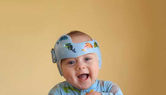
Endoscopic Surgery: This minimally invasive procedure is usually performed on infants younger than 6 months. Through small incisions, surgeons insert an endoscope to remove the fused sutures, allowing the skull to expand naturally as the child grows. The advantages of endoscopic surgery include smaller scars, shorter recovery times, and less overall risk compared to more invasive procedures. However, post-surgery, the infant will typically need to wear a specially molded helmet for several months to guide the skull into a normal shape as it grows.
Open Cranial Vault Remodeling: For older infants or when endoscopic surgery is not an option, open cranial vault remodeling is recommended. This procedure involves making larger incisions to directly reshape the affected portion of the skull. While this method is more invasive and involves a longer recovery period, it has a high success rate in correcting the skull shape and mitigating the risk of increased intracranial pressure.
Ideal Timing and Potential Risks: The timing for surgery is critical, with the best outcomes observed when the procedure is performed early in an infant’s life. Endoscopic surgery is typically performed when the child is between 1 to 6 months old, whereas open cranial vault remodeling can be done up to the age of 1 year or older. Risks associated with these surgeries include bleeding, infection, and the need for further surgeries, although these are relatively rare.
Non-surgical Interventions
In cases where craniosynostosis is mild or part of a watchful waiting strategy, non-surgical interventions may be applicable. These can include:
- Helmet Therapy: While not a treatment for the craniosynostosis itself, helmet therapy can be used after endoscopic surgery to help mold the skull into a more typical shape. It is also used in cases where craniosynostosis is suspected but not confirmed, to correct positional plagiocephaly, a condition that can mimic craniosynostosis.
- Physical Therapy: In some instances, physical therapy may be recommended to address developmental delays or motor skill challenges resulting from the craniosynostosis.
While surgical intervention remains the cornerstone of craniosynostosis treatment, understanding the full spectrum of options, including non-surgical approaches, allows for a more informed decision-making process. Early diagnosis and intervention are key to achieving the best possible outcomes for children with craniosynostosis, highlighting the importance of regular pediatric check-ups and monitoring.
Preparing for Treatment of Craniosynostosis
Here’s a comprehensive guide to help you navigate the journey from diagnosis to treatment, focusing on assembling the right healthcare team, understanding what to expect from consultations, and making pre-surgical preparations for both infants and their families.
Steps to Take Once a Diagnosis is Confirmed
1. Educate Yourself About the Condition: Learning about craniosynostosis is the first step. Understand the specific type your child has been diagnosed with, as there are several forms, each affecting the skull and the child’s development differently.
2. Seek Support: Connecting with support groups can provide emotional comfort and practical advice from other families who have gone through similar experiences.
Choosing the Right Healthcare Team
1. Find a Specialist: Look for a pediatric neurosurgeon or craniofacial surgeon with extensive experience in treating craniosynostosis. The right professional should not only have a proven track record but also make you feel comfortable and heard.
2. Multidisciplinary Approach: Opt for a hospital or center that offers a multidisciplinary team approach, including neurosurgeons, plastic surgeons, pediatricians, and support staff. This ensures all aspects of your child’s care are addressed.
3. What to Expect from Consultations: Initial consultations will involve detailed discussions about the diagnosis, treatment options (including the possibility of surgery), risks, and expected outcomes. Be prepared to ask questions and discuss any concerns you have about the procedure and recovery.
Pre-surgical Preparations for Infants and Their Families
1. Pre-surgical Testing: Your child may need to undergo several tests before surgery, such as imaging studies to assess the skull’s structure and brain function. Understanding these tests can help alleviate some anxieties about the process.
2. Discuss Anesthesia: An important part of the preparation involves discussions about anesthesia, as surgery for craniosynostosis is performed under general anesthesia. Anesthesiologists will explain how they keep your child comfortable and safe during the procedure.
3. Preparing Your Child: Although infants may not understand what is happening, older children might sense the anxiety of their parents. Keeping your emotions in check can help maintain a calm environment for your child.
4. Practical Arrangements: Make practical arrangements for the hospital stay and the recovery period at home. This includes organizing transportation, managing work and family commitments, and preparing your home for post-surgery care.
5. Emotional Support: It’s equally important to prepare emotionally. Speak with the healthcare team about what to expect in terms of recovery and any changes in your child’s appearance or behavior. Support from family, friends, and possibly a counselor can be invaluable during this time.
Preparing for the treatment of craniosynostosis involves thorough planning and emotional readiness. By choosing the right healthcare team, understanding the treatment process, and making both practical and emotional preparations, you can ensure the best possible outcome for your child and your family. Remember, you are not alone; support from healthcare professionals and fellow parents can provide guidance and reassurance throughout this journey.
Recovery and Aftercare of Craniosynostosis
Recovering from craniosynostosis surgery is a journey that requires patience, understanding, and the right resources. Families navigating this path will find that knowledge is their best ally. In this comprehensive guide, we delve into what to expect during the recovery period post-surgery, the importance of long-term follow-up care, and the support and resources available for families dealing with craniosynostosis.
What to Expect During the Recovery Period Post-Surgery
The immediate recovery period following craniosynostosis surgery is critical for the child’s healing and overall wellbeing. Typically, children may spend a few days in the hospital to allow healthcare professionals to closely monitor their recovery. Pain management is a key focus during this time, with medication often prescribed to ensure the child remains comfortable.
Swelling and bruising around the surgical area are common, but these symptoms gradually diminish over the following weeks. Parents and caregivers are advised to follow the surgeon’s aftercare instructions meticulously, which may include guidelines on how to properly position the child while sleeping, recommended activities, and signs of complications to watch for.
It’s also a time for emotional recovery. Families should be prepared for a range of emotions and understand that support from healthcare providers, counselors, or support groups can be incredibly beneficial.
Long-term Follow-up Care and Monitoring for Developmental Milestones
After the initial recovery phase, long-term follow-up care becomes essential. These follow-up visits allow the surgeon and other healthcare professionals to monitor the child’s skull growth, brain development, and overall health. Regular check-ups are crucial for identifying any potential issues early on and addressing them promptly.
Developmental monitoring is another critical aspect of long-term care. Children who have undergone craniosynostosis surgery may face challenges with cognitive, speech, and physical development. Early intervention programs and therapies, such as speech therapy or physical therapy, can play a significant role in supporting a child’s development.
Support and Resources for Families Dealing with Craniosynostosis
Facing craniosynostosis can feel overwhelming for families, but a wealth of support and resources are available. Many hospitals and healthcare providers offer access to support groups where families can share experiences and advice. These groups provide a sense of community and understanding that can be incredibly comforting.
Educational resources are also vital. Learning about craniosynostosis, understanding the surgical process, and knowing what to expect during recovery can empower families to advocate for their child’s needs effectively.
Moreover, nonprofit organizations dedicated to craniosynostosis offer additional resources, including financial assistance, educational materials, and connections to specialists in the field. Engaging with these organizations can provide families with the support they need to navigate the challenges of craniosynostosis.
Innovations in Craniosynostosis Treatment
In recent years, the medical field has witnessed groundbreaking advancements in the surgical treatment of craniosynostosis, enhancing patient outcomes and transforming the approach to care. This article delves into the latest innovations in craniosynostosis treatment, highlighting the pivotal role of 3D printing and custom implants, and explores promising future directions in research and treatment options.
Recent Advancements in the Surgical Treatment of Craniosynostosis
The surgical landscape for craniosynostosis has evolved significantly, moving from traditional, more invasive procedures to minimally invasive techniques. These advancements not only reduce the risks associated with surgery but also shorten recovery times, making the process less daunting for patients and their families. Among these, endoscopic surgery stands out as a less invasive option that allows for quicker recovery and less noticeable scarring, marking a significant step forward in the field.
The Role of 3D Printing and Custom Implants in Improving Outcomes
One of the most transformative innovations in the treatment of craniosynostosis is the integration of 3D printing technology. This technology facilitates the creation of custom-made implants and surgical guides tailored to the unique anatomy of each patient. By providing surgeons with precise tools and implants that perfectly fit the patient’s cranial structure, 3D printing significantly enhances surgical accuracy, reduces operation times, and improves overall outcomes. The ability to plan surgeries with unparalleled precision has not only improved the aesthetic results but also minimized potential complications, making it a game-changer in craniosynostosis surgery.
Future Directions in Research and Treatment Options
Looking ahead, the future of craniosynostosis treatment holds immense promise. Researchers are exploring novel materials for implants that mimic the properties of natural bone, encouraging bone regeneration and reducing the likelihood of rejection. Additionally, advances in gene therapy and molecular biology may offer less invasive treatment alternatives, targeting the genetic and molecular underpinnings of craniosynostosis. As understanding of the condition deepens, personalized medicine approaches that tailor treatments to the genetic profile of each patient could become a reality, offering hope for even better outcomes.
However, the innovations in craniosynostosis treatment, particularly the use of 3D printing and custom implants, have revolutionized patient care, offering safer, more effective, and less invasive options. As research continues to advance, the future of treatment looks bright, with the potential for even more personalized and precise interventions that could further improve the quality of life for patients with craniosynostosis. The ongoing commitment to innovation and research in this field underscores a promising horizon for treatment options, ensuring that affected individuals have access to the best possible care.
FAQs on Craniosynostosis for Parents
What is Craniosynostosis?
Craniosynostosis is a condition where the sutures in a baby’s skull close too early, preventing the skull from growing properly and potentially affecting brain development. This premature fusion can lead to changes in the shape of the head and facial features.
How Common is Craniosynostosis?
Craniosynostosis affects approximately 1 in every 2,500 births. It can occur as an isolated condition or as part of a syndrome involving other health issues.
What Causes Craniosynostosis?
The exact cause of craniosynostosis is often unknown, but it can be attributed to a combination of genetic and environmental factors. In some cases, it’s associated with certain genetic syndromes.
How is Craniosynostosis Diagnosed?
Craniosynostosis is typically diagnosed through physical examination and imaging tests such as X-rays or CT scans. These tests help determine which sutures have fused and assess the impact on the skull and brain.
What are the Symptoms of Craniosynostosis?
Symptoms can vary depending on which sutures are affected but often include a misshapen skull, a hard ridge along the sutures, and slow or no growth in the head size as the baby grows. In severe cases, increased pressure within the skull can lead to developmental delays or cognitive issues.
How is Craniosynostosis Treated?
Treatment usually involves surgery to correct the shape of the skull and allow for normal brain growth. The type of surgery depends on the severity of the condition and the sutures affected. Post-surgery, follow-up care is crucial to monitor the child’s development.
Can Craniosynostosis Lead to Complications?
If left untreated, craniosynostosis can lead to increased intracranial pressure, developmental delays, and visual impairments. However, with timely and appropriate treatment, most children go on to live healthy lives.
Is There a Way to Prevent Craniosynostosis?
Since the exact cause of craniosynostosis is not fully understood and often involves genetic factors, there is no known way to prevent it. However, accessing prenatal care and following a healthy lifestyle during pregnancy can help reduce the risk of congenital conditions.
Where Can I Find Support?
Caring for a child with craniosynostosis can be challenging. Support from healthcare professionals, including pediatricians, neurosurgeons, and genetic counselors, is vital. Additionally, joining support groups can connect you with other families navigating similar experiences.
Conclusion:
For parents and caregivers, being vigilant about the symptoms and seeking medical advice without delay can make a substantial difference in the treatment journey. Consulting with specialists, such as pediatric neurosurgeons or craniofacial experts, is essential to assess the condition accurately and determine the most suitable intervention. These professionals are equipped with the knowledge and skills to offer tailored treatment plans that cater to the unique needs of each child, ensuring the best possible outcomes.
Encouragement cannot be overstated for parents navigating this challenging situation. The road may seem daunting at first, but early intervention and specialized care pave the way for promising results. Through prompt action and expert guidance, you can support your child’s health and well-being, laying a strong foundation for their future growth and development.
Remember, you are not alone in this journey. A multidisciplinary team of healthcare providers will be by your side, offering support, expertise, and compassion every step of the way. By recognizing the signs of craniosynostosis early and seeking specialized care, you are taking a proactive step towards ensuring a healthier, brighter future for your child.